Imaging often becomes the first step when patients report persistent coughing or unexplained shortness of breath. When physical exams and patient history offer limited clarity, radiology becomes essential. Doctors look for structural changes in the lungs, often invisible through stethoscope or symptoms alone. Not all lung diseases present with loud or dramatic signs. Subtle infiltrates, mild fibrosis, or hidden nodules often escape detection without imaging. For patients with nonspecific symptoms like fatigue or dry cough, imaging helps decide what to investigate further. The decision to order a scan depends on the clinical picture and duration of complaints. A standard chest X-ray is usually the first imaging tool used.
Chest X-rays provide a quick overview of lung structure but may miss smaller or hidden issues
Chest X-rays provide a quick overview of lung structure but may miss smaller or hidden issues. This method shows lung fields, heart size, diaphragm shape, and pleural spaces. It can detect large masses, fluid buildup, pneumonia, and collapsed lung segments. However, smaller abnormalities like early tumors or interstitial changes may not appear clearly. Overlapping structures and limited resolution affect accuracy, especially in early or mild cases. Doctors often compare with prior films, looking for changes over time. In emergency settings, chest X-rays guide immediate decisions. Still, when results are unclear, further imaging is often required. X-rays show the forest, not always the tree.
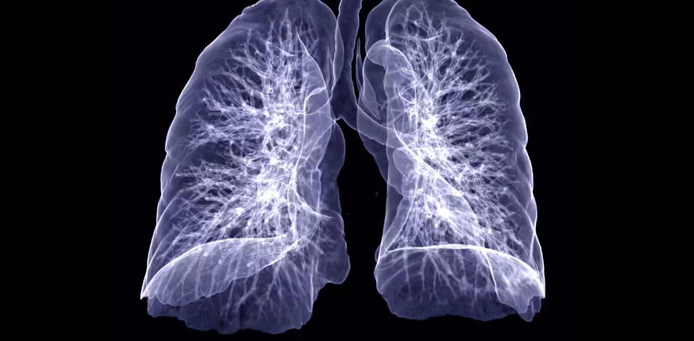
Computed tomography gives more detailed images, often revealing patterns not seen in standard radiographs
Computed tomography gives more detailed images, often revealing patterns not seen in standard radiographs. CT scans produce cross-sectional views of lung tissue, offering a clearer map of anatomy. This method is highly sensitive for detecting nodules, scarring, and vascular abnormalities. It plays a key role in diagnosing diseases like pulmonary embolism, interstitial lung disease, or cancer. Radiologists assess density, distribution, and structure of lesions to narrow down possibilities. For instance, ground-glass opacities might suggest inflammation or infection. Honeycombing could point to fibrosis. These visual patterns create diagnostic direction. CT imaging bridges the gap between suspicion and specificity, guiding biopsy or treatment.
High-resolution CT is often used when interstitial or fibrotic lung disease is suspected
High-resolution CT is often used when interstitial or fibrotic lung disease is suspected. Unlike standard CT, it uses thin slices for greater detail in the lung parenchyma. This technique helps identify subtle architectural changes not visible with thicker cuts. Doctors use HRCT to classify fibrosis type, evaluate extent, and detect progression. Distribution matters—whether it’s upper, lower, peripheral, or central. These patterns often align with known diseases like idiopathic pulmonary fibrosis or sarcoidosis. HRCT helps differentiate between active inflammation and irreversible scarring. This distinction shapes treatment decisions, especially when steroids or immunosuppressants are considered. Serial HRCT scans monitor change over time, revealing response or resistance.
CT pulmonary angiography is preferred when pulmonary embolism is strongly suspected
CT pulmonary angiography is preferred when pulmonary embolism is strongly suspected. This imaging technique visualizes the pulmonary arteries after contrast injection. It shows blockages, narrowing, or sudden stops in blood flow. Shortness of breath, chest pain, and low oxygen levels often lead to this test. CTPA is fast, detailed, and widely available in emergency departments. It replaced older methods like V/Q scanning in most acute cases. A negative result can rule out embolism with high confidence. However, patients must be screened for contrast allergy and kidney function beforehand. When done promptly, this scan can prevent delay in critical treatment decisions.
Positron emission tomography helps distinguish between benign and malignant lung nodules
Positron emission tomography helps distinguish between benign and malignant lung nodules. PET scans measure metabolic activity in tissue using radioactive tracers. Cancerous cells often absorb more tracer due to high glucose use. If a lung nodule lights up strongly, malignancy is more likely. However, infection and inflammation can also cause uptake. That’s why PET is interpreted alongside CT or biopsy results. This tool is often used after initial detection to guide further steps. It helps determine whether surgery, observation, or biopsy is most appropriate. PET imaging doesn’t diagnose cancer alone, but it increases diagnostic certainty.
Ultrasound is used to evaluate pleural effusions or guide procedures like thoracentesis
Ultrasound is used to evaluate pleural effusions or guide procedures like thoracentesis. Unlike X-ray or CT, it doesn’t visualize the lung parenchyma well. But it excels at identifying fluid, guiding safe drainage, and monitoring response to therapy. It can differentiate between simple and complex effusions. Ultrasound is also portable, radiation-free, and usable at the bedside. Emergency and intensive care doctors often rely on it for real-time assessment. During thoracentesis, ultrasound ensures the needle avoids lung tissue and punctures the fluid pocket directly. For patients unable to undergo CT, ultrasound provides a safer option in certain contexts.
MRI is less commonly used but valuable for specific cases involving the chest wall or heart-lung interface
MRI is less commonly used but valuable for specific cases involving the chest wall or heart-lung interface. Its ability to show soft tissue makes it suitable for evaluating tumors extending into muscle, nerve, or spine. It’s also useful in pediatric or pregnant patients who need to avoid radiation. In cases where CT findings are inconclusive, MRI may provide additional clarity. It can assess blood flow without contrast in patients with kidney issues. However, limitations include motion artifacts and longer scan times. Lung tissue contains air, which reduces MRI effectiveness. Still, for certain complex or sensitive cases, MRI has a unique role in lung imaging.
Fluoroscopy and ventilation-perfusion scans still play a role in specific lung evaluations
Fluoroscopy and ventilation-perfusion scans still play a role in specific lung evaluations. Fluoroscopy allows dynamic observation of diaphragmatic motion or airway collapse. It helps diagnose vocal cord paralysis or tracheomalacia. V/Q scans measure air and blood flow in the lungs. They’re useful for detecting chronic embolism, especially when CT is contraindicated. In patients with allergy to contrast dye, V/Q scanning becomes the safer choice. Though older, these tests retain relevance in certain contexts. They provide functional data that static images sometimes miss. Their value lies not in replacement but in complementing newer techniques.
Final diagnosis rarely depends on one image but a combination of patterns, symptoms, and clinical context
Final diagnosis rarely depends on one image but a combination of patterns, symptoms, and clinical context. Imaging shows structure, not always meaning. A shadow on CT may mean infection, tumor, or artifact. Doctors correlate findings with physical exams, blood tests, and patient history. They consider age, risk factors, exposures, and preexisting conditions. One scan may raise suspicion, but only in context does it become actionable. Biopsy or bronchoscopy may follow. In lung medicine, certainty is built layer by layer. Imaging leads the path, but interpretation walks it.